Introduction
The shoulder joint is a complex structure that enables a wide range of movements and plays a crucial role in our daily activities. Understanding the anatomy of the shoulder joint is essential for diagnosing and treating shoulder-related issues. In this article, we will explore the key components of the shoulder joint and their functions.
Bones of the Shoulder Joint
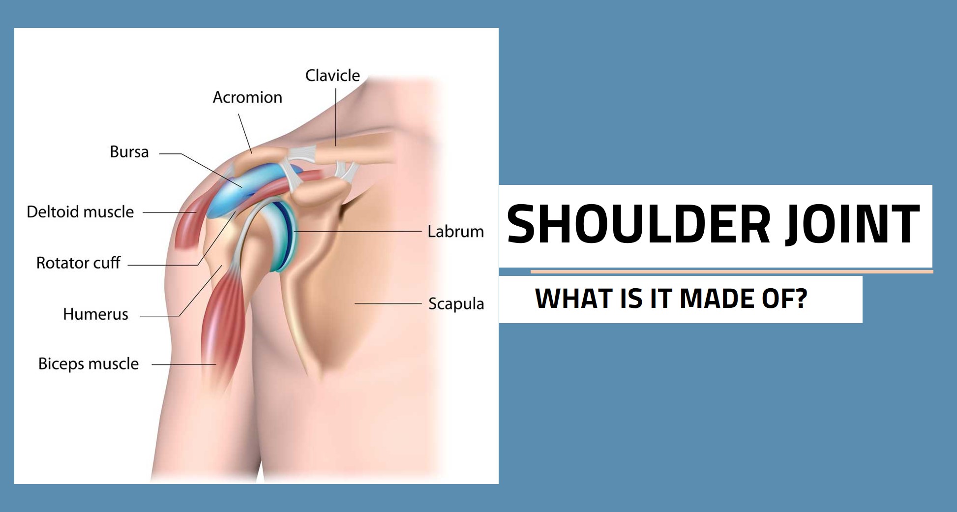
The shoulder joint consists of three main bones: the humerus (upper arm bone), the scapula (shoulder blade), and the clavicle (collarbone). The rounded head of the humerus fits into the shallow socket of the scapula, forming the main ball-and-socket joint of the shoulder. The clavicle connects the sternum (breastbone) to the scapula, providing stability and support to the shoulder joint.
Articular Cartilage
Articular cartilage covers the ends of the bones in the shoulder joint. It is a smooth, slippery tissue that helps reduce friction and allows for smooth movement between the bones. The cartilage absorbs shock and provides cushioning, protecting the bones from damage during movement.
Ligaments
Ligaments are tough, fibrous bands of connective tissue that help stabilise the shoulder joint. The main ligaments in the shoulder joint include the glenohumeral ligaments, which surround the joint and reinforce its capsule, and the coracoclavicular ligaments, which connect the clavicle to the scapula. These ligaments play a crucial role in maintaining the integrity of the joint and preventing excessive movement that could lead to injury.
Muscles
Several muscles surround the shoulder joint, enabling its wide range of movements. The rotator cuff muscles, including the supraspinatus, infraspinatus, teres minor, and subscapularis, are a group of muscles that originate from the scapula and attach to the humerus. These muscles work together to stabilize the joint and initiate shoulder movements. Other important muscles include the deltoid, which covers the shoulder joint and aids in arm abduction, and the pectoralis major and latissimus dorsi, which contribute to movements involving the shoulder joint.
Bursae
Bursae are small fluid-filled sacs located between bones, tendons, and muscles. They help reduce friction and provide cushioning during movement. In the shoulder joint, two main bursae are present: the subacromial bursa, located beneath the acromion process of the scapula, and the subscapular bursa, positioned between the subscapularis muscle and the scapula. These bursae protect the tendons and muscles from rubbing against bone during shoulder movements.
Understanding the anatomy of the shoulder joint is essential for diagnosing and managing shoulder-related conditions. The bones, articular cartilage, ligaments, muscles, and bursae work together to ensure the stability and mobility of the shoulder joint. By recognising the key components and their functions, patients can understand their shoulder-related issues better.