The knee joint is a crucial weight-bearing joint in the human body that facilitates movement and stability. Understanding its complex anatomy is essential for comprehending its function and identifying potential sources of pain or injury. In this article, we will explore the anatomy of the knee joint in detail.
Structure of the Knee Joint
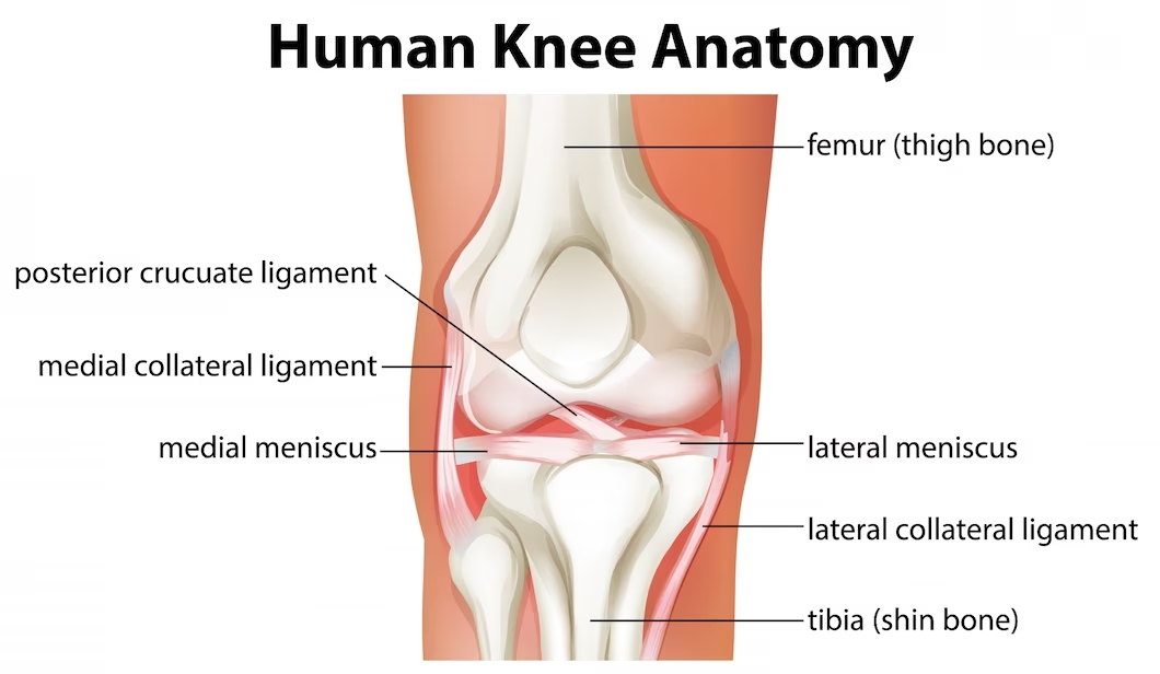
The knee joint is a synovial hinge joint that connects the femur (thigh bone) to the tibia (shin bone). It consists of three main components: bones, ligaments, and cartilage. The femur’s rounded ends, known as condyles, articulate with the flat upper surface of the tibia, forming the primary joint. The patella (kneecap), a small bone, lies in front of the joint and provides protection.
Ligaments
The knee joint is stabilized by four major ligaments. The medial collateral ligament (MCL) and lateral collateral ligament (LCL) are located on the sides of the knee, preventing sideways motion. The anterior cruciate ligament (ACL) and posterior cruciate ligament (PCL) are situated inside the joint. The ACL prevents the tibia from sliding forward, while the PCL prevents backward movement.
Menisci
The knee joint contains two menisci, which are C-shaped pieces of fibrocartilage located between the femur and tibia. The medial meniscus is positioned on the inner side, and the lateral meniscus is on the outer side. They act as shock absorbers, providing cushioning and distributing forces during movement.
Articular Cartilage
The surfaces of the bones within the knee joint are covered with a layer of smooth articular cartilage. This cartilage reduces friction between the bones, allowing smooth gliding during movement. It also helps absorb shock and evenly distribute loads across the joint.
Bursae
The knee joint contains several small fluid-filled sacs called bursae. These bursae reduce friction and provide cushioning between tendons, ligaments, and bones. Bursitis, an inflammation of these sacs, can occur due to overuse or injury.
Muscles
Several muscles surround and support the knee joint. The quadriceps muscles, located at the front of the thigh, help in knee extension. The hamstrings, located at the back of the thigh, assist in knee flexion. Other muscles, such as the calf muscles and the iliotibial band (IT band), also play a role in knee stability and movement.
Understanding the anatomy of the knee joint is vital for understanding knee-related issues. Proper care and maintenance can help promote joint health and function.
Source/s: Banner image by Freepik